

Interactive 3D
Turn and open each kind of tooth layer by layer, see where a bite load goes, and watch a restoration seat on its preparation.
Tooth explorer
Pick a tooth, turn it, open it and switch its layers. Tap a label to learn what it is.

Central incisor. A front tooth for cutting, with a thin incisal edge. The upper central incisor usually has one root.
Lateral incisor. Smaller than the central incisor and also for cutting. Usually one root.
Canine. A single pointed cusp for tearing. Usually one root.
First premolar. Two cusps, buccal and lingual, for grinding. The upper first premolar usually has two roots, buccal and palatal.
First molar. Four main cusps for grinding. The upper first molar usually has three roots: mesiobuccal, distobuccal and palatal.
Enamel
The hardest and most mineralized tissue in the body, about 96% mineral by weight. It covers the crown.
Dentinoenamel junction
The boundary where enamel meets dentin. Turn it on to see the dentin surface under the enamel.
Dentin
The most plentiful tissue of the tooth: under the enamel in the crown and under the cementum on the root.
Pulp chamber
Soft tissue in the center of the crown, with nerves and blood vessels.
Root canals
The pulp continues through each root in its root canal.
Cementum and PDL
Cementum covers the root. The periodontal ligament (PDL) attaches it to the alveolar bone.
Tap a label on the model, or a layer, to read about it.
Keyboard: with the model focused, Left and Right turn it, Up and Down tilt it, plus and minus zoom, 0 resets the view.
Learn more
Where the bite load goes
Press and drag on the tooth to load it, then compare an intact tooth, an MOD cavity and a bonded porcelain inlay.
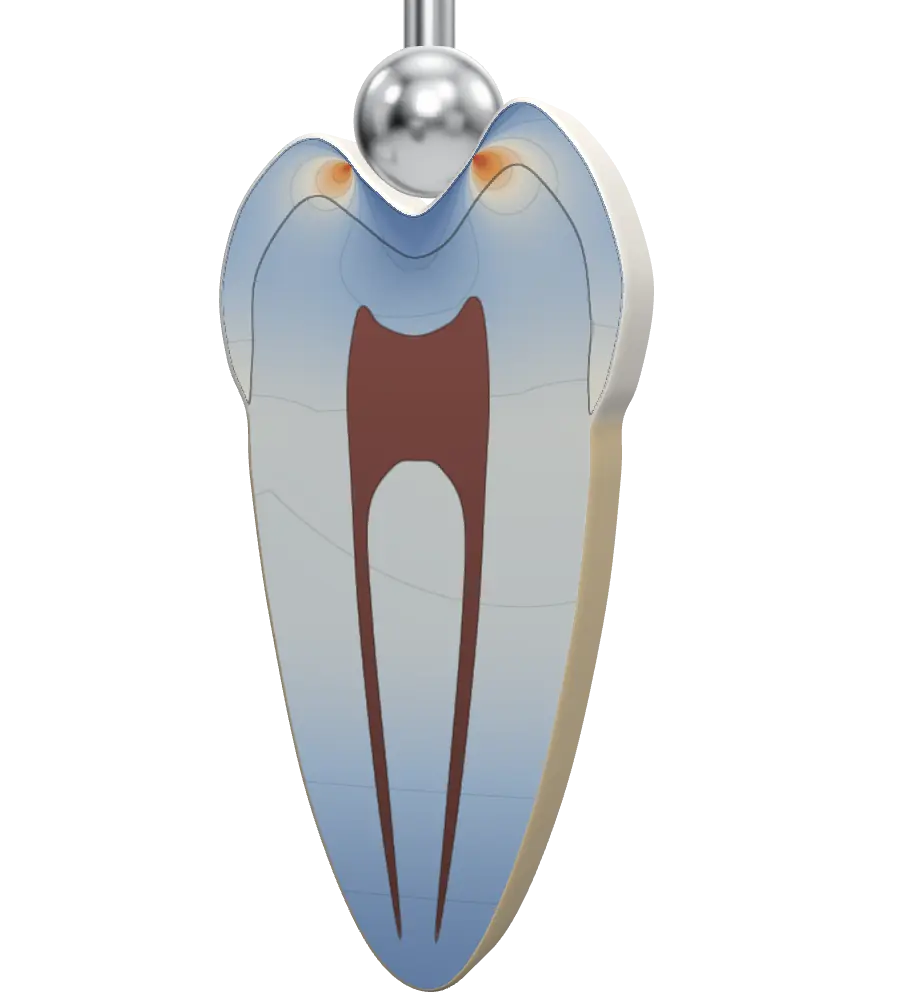
Intact tooth. Under the bite, the cusps barely move: 0.4 µm of cusp widening at 100 N.1
MOD cavity. With the center of the crown removed, the same bite pushes the cusps apart: 9 to 12 µm of cusp widening with MO and MOD cavities.1
Bonded porcelain inlay. Bonded in place, it holds the cusps together again: 0.4 µm, full recovery of cusp stiffness.1
Figure 1. A lower molar in cross section under a bite load: intact, with an MOD cavity, and restored with a bonded porcelain inlay. An illustration of the biomimetic principle, based on the research of Dr. Pascal Magne.2 Colors are illustrative, not measured stress values, and cusp movement is exaggerated. Cusp widening values are from Dr. Magne’s micro-CT finite element model.1
- Magne P. Efficient 3D finite element analysis of dental restorative procedures using micro-CT data. Dent Mater. 2007;23(5):539-548. doi:10.1016/j.dental.2006.03.013
- Magne P, Belser U. Bonded Porcelain Restorations in the Anterior Dentition: A Biomimetic Approach. Chicago: Quintessence Publishing; 2002.
A restoration on its preparation
Slide to seat a bonded porcelain veneer on a front tooth, or an onlay on a molar. Drag the model to turn it.
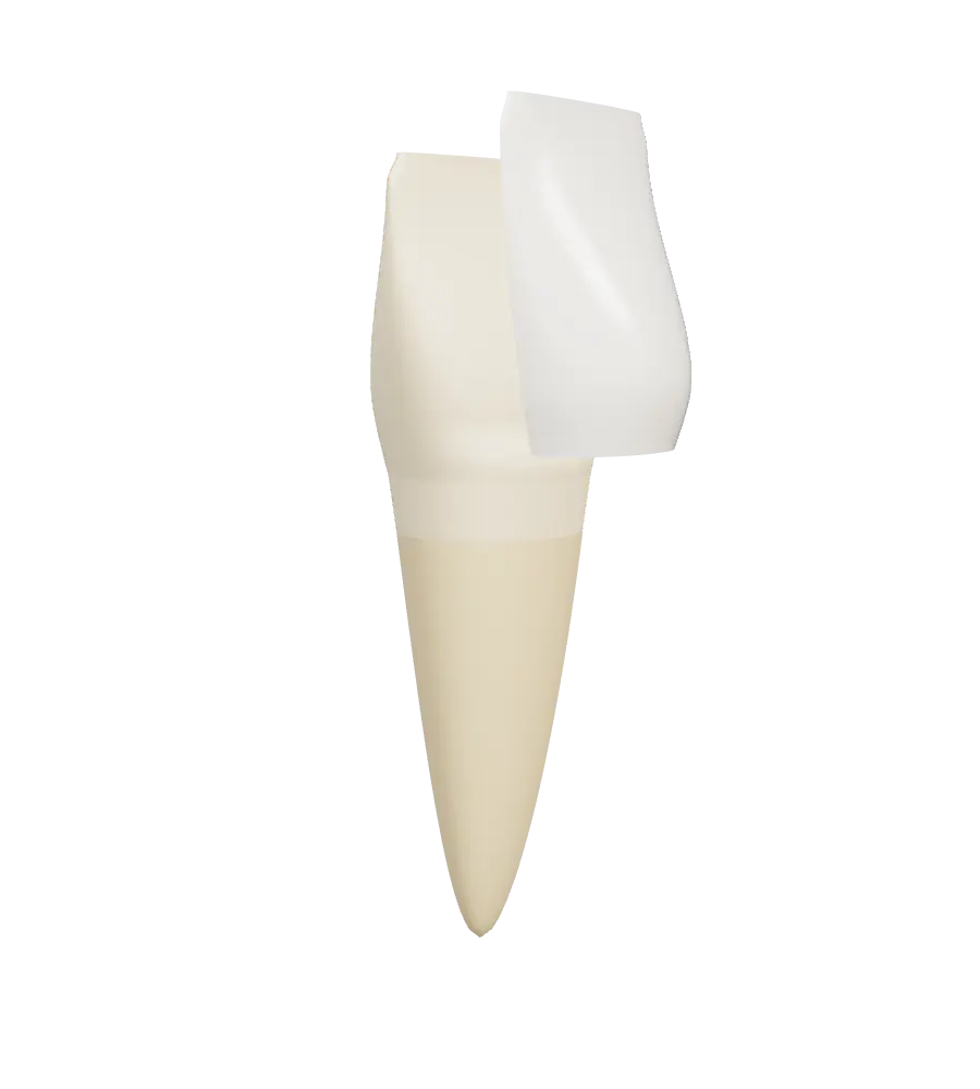
Porcelain veneer. A bonded porcelain veneer covers the labial (front) surface and the incisal edge of an anterior tooth.
Onlay. An onlay restores the chewing surface of a back tooth and covers one or more of its cusps.
Illustrative shapes, not a clinical preparation guide.
Learn more
Sources and credits
Anatomy descriptions are general dental anatomy from these references:
- Morris AL, Tadi P. Anatomy, Head and Neck, Teeth. In: StatPearls. Treasure Island (FL): StatPearls Publishing. NCBI Bookshelf NBK557543.
- Farci F, Soni A. Histology, Tooth. In: StatPearls. Treasure Island (FL): StatPearls Publishing. NCBI Bookshelf NBK572055.
- Torabi S, Soni A. Histology, Periodontium. In: StatPearls. Treasure Island (FL): StatPearls Publishing. NCBI Bookshelf NBK570604.
- Nelson SJ. Wheeler’s Dental Anatomy, Physiology and Occlusion. 11th ed. Elsevier; 2020.
- Magne P. Efficient 3D finite element analysis of dental restorative procedures using micro-CT data. Dent Mater. 2007;23(5):539-548. doi:10.1016/j.dental.2006.03.013
Every 3D model on this page is procedural: built in code, with no third party models or images. Shapes are simplified for teaching and are not to clinical scale.